Reporter Genes I
Patent Pending
8 Curated Reporter genes with varying use cases - ranging from in vitro work to in vivo imaging.
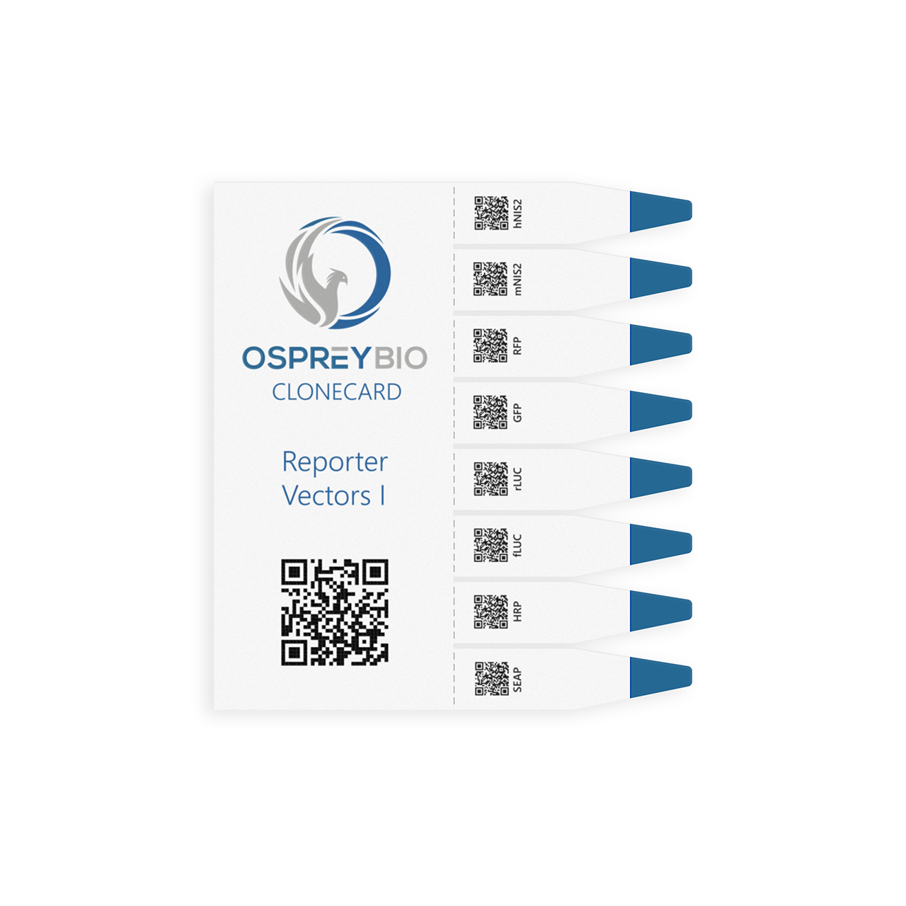
Genes in Collection I contain SEAP, HRP, fLUC, rLUC, GFP, RFP, mNIS2, hNIS2.
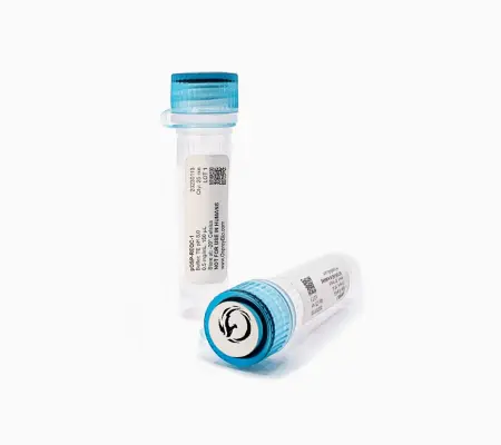
- Ready-to-use plasmid after just one elution (~1 hour)
- Safe Long Term Storage:
Can be stored flat at ambient temperature without the need for uninterrupted electricity supply (as in freezer storage) or the need for isolation from harmful agents (such as metal ions)
Vectors Included with this Card
Vector 1
SEAP (Secreted Alkaline Phosphatase)
Functionality
In its function as a reporter, SEAP (Secreted Alkaline Phosphatase) is a non-invasive, secretion signal with many applications in both the medical and biotechnology field.
Why this vector?
An easy to use and measure reporter, SEAP's many use cases make it a great choice for our system.
Use Cases
SEAP has been widely used as a secreted serum reporter for accurate long-term monitoring of gene expression.
- Abruzzese RV, Godin D, Burcin M, Mehta V, French M, Li Y, O’Malley BW, Nordstrom JL. “Ligand-dependent regulation of plasmid-based transgene expression in vivo.” Hum Gene Ther. 1999;10:1499–1507.
- Blacklock J, You YZ, Zhou QH, Mao G, Oupicky D. “Gene delivery in vitro and in vivo from bioreducible multilayered polyelectrolyte films of plasmid DNA.” Biomaterials. 2009;30:939–950.
- Brown PA, Khan AS, Draghia-Akli R. “Delivery of DNA into skeletal muscle in large animals.”Methods Mol Biol. 2008;423:215–224.
- Yang TT, Sinai P, Kitts PA, Kain SR. “Quantification of gene expression with a secreted alkaline phosphatase reporter system.” Biotechniques. 1997 Dec;23(6):1110-4. doi: 10.2144/97236pf01. PMID: 9421645.
Vector 2
HRP (Horseradish Peroxidase)
Functionality
Horseradish Peroxidase (HRP) is useful for many bioassays and has been used for electron microscopy as a transgene. HRP has the ability to catalyze the transfer of two electrons from a substrate to hydrogen peroxide via a two-electron oxidation of HRP event to generate water and an oxidized substrate. HRP is a popular detection label used for protein target detection, and is the most popular enzyme used in western blotting. It is also commonly used in immunohistochemistry and ELISA because it generates colored compounds. In photometric assays, HRP reacts with a substrate to produce chemiluminescent, chromogenic, or fluorescent signals upon oxidation, and it can amplify a weak signal from a target molecule.
HRP has several advantages over other reporters due to its combination of beneficial traits. HRP is robust, inexpensive, relatively small (allowing for more effective penetration into cells without risking interference with the conjugated protein function), readily binds to antibodies in an active form, oxidizes a wide range of substrates to produce optical signals, and more. The other key feature about HRP is that it has a high rate of turnover and this results in an efficient reaction, producing an abundance of reactant products in a short span of time at normal pH.
Note: Osprey Bio’s HRP does not have a subcellular localization signal.
Why this vector?
Osprey Bio’s research indicated that HRP was one of the top used reporter genes in gene & cell therapy research due to its high sensitivity, simplicity, and wide usage applications.
Use Cases
- Berchmans, Sheela, et al. "PAMAM dendrimer modified reduced graphene oxide postfunctionalized by horseradish peroxidase for biosensing H2O2." Methods in Enzymology. Vol. 609. Academic Press, 2018. 143-170.
- Hopkins C, Gibson A, Stinchcombe J, Futter C. “Chimeric molecules employing horseradish peroxidase as reporter enzyme for protein localization in the electron microscope.” Methods Enzymol. 2000;327:35-45. doi: 10.1016/s0076-6879(00)27265-0. PMID: 11044972.
- Hu, S., Q. Lu, and Y. Xu. "Biosensors based on direct electron transfer of protein." Electrochemical Sensors, Biosensors and their Biomedical Applications (2008): 531.
- Kalluri, Ankarao, et al. "Exfoliated and water dispersible biocarbon nanotubes for enzymology applications." Methods in Enzymology. Vol. 630. Academic Press, 2020. 407-430.
- Schikorski T, Young SM Jr, Hu Y. “Horseradish peroxidase cDNA as a marker for electron microscopy in neurons.” J Neurosci Methods. 2007 Sep 30;165(2):210-5. doi: 10.1016/j.jneumeth.2007.06.004. Epub 2007 Jun 12. PMID: 17631969.
- Sharma, Bhupesh, Kanishk Luhach, and G. T. Kulkarni. "In vitro and in vivo models of BBB to evaluate brain targeting drug delivery." Brain targeted drug delivery system. Academic Press, 2019. 53-101.
Vector 3
fLUC (Firefly Luciferase)
Functionality
Firefly Luciferase (fLuc) catalyzes a two-step reaction in which oxidation of D-luciferin yields green to yellow light. Incorporating Coenzyme A (CoA) optimizes the bioluminescent reaction to yield maximal luminescence intensity that decays slowly over several minutes. The influence of CoA on fLuc generates a relatively stable and sensitive luminescence in less that 0.3 seconds.
fLuc is a convenient-to-use monomer that becomes a mature enzyme directly upon translation of its mRNA, and demonstrates catalytic activity immediately after release from the ribosome. Its relatively short half-life compared to other commonly used reporters also makes it suitable for reporting changes in gene expression over a short period of time.
Why this vector?
fLuc is the most commonly used bioluminescent reporter. The broad usage of firefly luciferase as a genetic reporter is due to its high sensitivity and convenience of use, noninvasive nature, and the tight coupling of protein synthesis with enzyme activity.
Use Cases
- Close DM, Xu T, Sayler GS, Ripp S. “In vivo bioluminescent imaging (BLI): noninvasive visualization and interrogation of biological processes in living animals.” Sensors (Basel). 2011;11(1):180-206. doi: 10.3390/s110100180. Epub 2010 Dec 28. PMID: 22346573; PMCID: PMC3274065.
- Fan, Xiujun & Nayak, Nihar. (2014). “Lentivirus Transduction of Gene Tags in Blastocysts.” 10.1016/B978-0-12-394445-0.00040-0.
- Neefjes, M., Housmans, B.A.C., van den Akker, G.G.H. et al. “Reporter gene comparison demonstrates interference of complex body fluids with secreted luciferase activity.” Sci Rep 11, 1359 (2021). https://doi.org/10.1038/s41598-020-80451-6
- Schoene C, Bennett SP, Howarth M. SpyRings Declassified: A Blueprint for Using Isopeptide-Mediated Cyclization to Enhance Enzyme Thermal Resilience. Methods Enzymol. 2016;580:149-67. doi: 10.1016/bs.mie.2016.05.004. Epub 2016 Jun 16. PMID: 27586332.
Vector 4
rLUC (Renilla Luciferase)
Functionality
Renilla luciferase (rLuc) is a bioluminescent enzyme that is popular for its wide range of applications and its ability to catalyze a blue-green bioluminescent reaction upon mechanical stimulation using only molecular oxygen. It is generally used as a reporter vector to assess transcriptional expression, and has been the subject of several improvements over the years. Due to these improvements, rLuc can now be quantitated in living cells in situ or in vivo, and no longer faces the issues of high background and reduced assay sensitivity typical to autoluminescence.
Why this vector?
rLuc is one of the three most commonly used bioluminescent reporters and provides many of the same benefits as the most commonly used bioluminescent reporter, firefly luciferase. It has been extensively studied and used as a reporter vector.
Use Cases
- Abruzzese RV, Godin D, Burcin M, Mehta V, French M, Li Y, O’Malley BW, Nordstrom JL. “Ligand-dependent regulation of plasmid-based transgene expression in vivo.” Hum Gene Ther. 1999;10:1499–1507.
- Blacklock J, You YZ, Zhou QH, Mao G, Oupicky D. “Gene delivery in vitro and in vivo from bioreducible multilayered polyelectrolyte films of plasmid DNA.” Biomaterials. 2009;30:939–950.
- Brown PA, Khan AS, Draghia-Akli R. “Delivery of DNA into skeletal muscle in large animals.” Methods Mol Biol. 2008;423:215–224.
- Yang TT, Sinai P, Kitts PA, Kain SR. “Quantification of gene expression with a secreted alkaline phosphatase reporter system.” Biotechniques. 1997 Dec;23(6):1110-4. doi: 10.2144/97236pf01. PMID: 9421645.
Vector 5
EGFP (Enhanced Green Fluorescent Protein)
Functionality
The Open Reading Frame (ORF) encoding Enhanced Green Fluorescent Protein (EGFP) is a useful tool for investigating numerous cellular activities. The most basic application is to define if a pDNA-based expression vector was successfully introduced into the nuclei of transfected cells. Additionally, the intensity of fluorescence can be used in select imaging systems and/or cell sorting devices to evaluate the relative strength of promoters. EGFP has also been used widely to develop transgenic animals, advanced biosensors, and to track the efficiency of gene delivery systems.
Why this vector?
EGFP is one of the most widely used fluorescent protein reporters within the biotech research community making it an essential gene in any researcher's toolkit.
Use Cases
EGFP has been widely used in cell culture, transgenic animals, and for gene delivery.
- K-M R Prasad, Y Xu, Z Yang, S T Acton, B A French "Robust cardiomyocyte-specific gene expression following systemic injection of AAV: in vivo gene delivery follows a Poisson distribution"
- Zicong Xie, Ruize Sun, Chunyun Qi, Shuyu Jiao, Yuan Jiang, Zhenying Liu, Dehua Zhao, Ruonan Liu, Qirong Li, Kang Yang, Lanxin Hu, Xinping Wang, Xiaochun Tang, Hongsheng Ouyang, Daxin Pang "Generation of a pHSPA6 gene-based multifunctional live cell sensor"
- Yongjie Yang, Svetlana Vidensky, Lin Jin, Chunfa Jie, Ileana Lorenzini, Miriam Frankl, Jeffrey D Rothstein Molecular comparison of GLT1+ and ALDH1L1+ astrocytes in vivo in astroglial reporter mice
Vector 6
RFP (Red Fluorescent Protein)
Functionality
The Open Reading Frame (ORF) encoding Red Fluorescent Protein (plobRFP) is a useful tool for investigating numerous cellular activities. The most basic application is to define if a pDNA-based expression vector was successfully introduced into the nuclei of transfected cells. Additionally, the intensity of fluorescence can be used in select imaging systems and/or cell sorting devices to evaluate the relative strength of promoters. EGFP has also been used widely to develop transgenic animals, advanced biosensors, and to track the efficiency of gene delivery systems.
Why this vector?
plobRFP, derived from the coral special Porites lobata, displays a far red-shifted emission signal. Though not as characterized as some RFPs, plobRFP does not have IP constraints. Supporting data is available at: https://pubmed.ncbi.nlm.nih.gov/31853751
Vector 7
mNIS2 (Sodium Iodide Symporter)
Functionality
In its function as a reporter, the Sodium Iodide Symporter (NIS) is a non-invasive, precise imaging reporter gene with many applications in both the medical and biotechnology field. It enables researchers to track cells in vivo via nuclear imaging due to its ability to drive the uptake of radioactive iodine into cells as well as several other radiotracers at the site of transgene expression.
Why this vector?
NIS is the most common human reporter gene in use, and NIS imaging has several key advantages: it produces high-resolution 3D images, it can track anatomical gene therapy delivery with a high degree of precision, it is translational from small to large animals as well as humans, and it is non-immunogenic when species-specific and therefore usable in longitudinal imaging studies. Because NIS can be used for generating transgenic animals, generating GM-Cells, and being delivered directly to animals and/or humans, it’s essential that the proper gene from the appropriate species be used so there is no immune rejection. Therefore, the pOSP-CCRV-1 includes both mNIS2 and hNIS2.
Use Cases
- Boutagy NE, Ravera S, Papademetris X, Onofrey JA, Zhuang ZW, Wu J, Feher A, Stacy MR, French BA, Annex BH, Carrasco N, Sinusas AJ. Noninvasive In Vivo Quantification of Adeno-Associated Virus Serotype 9-Mediated Expression of the Sodium/Iodide Symporter Under Hindlimb Ischemia and Neuraminidase Desialylation in Skeletal Muscle Using Single-Photon Emission Computed Tomography/Computed Tomography. Circ Cardiovasc Imaging. 2019 Jul;12(7):e009063. doi: 10.1161/CIRCIMAGING.119.009063. Epub 2019 Jul 12. PMID: 31296047; PMCID: PMC6629470.
- Ravera S, Reyna-Neyra A, Ferrandino G, Amzel LM, Carrasco N. The Sodium/Iodide Symporter (NIS): Molecular Physiology and Preclinical and Clinical Applications. Annu Rev Physiol. 2017 Feb 10;79:261-289. doi: 10.1146/annurev-physiol-022516-034125. PMID: 28192058; PMCID: PMC5739519.
Vector 8
hNIS2 (Sodium Iodide Symporter)
Functionality
In its function as a reporter, the Sodium Iodide Symporter (NIS) is a non-invasive, precise imaging reporter gene with many applications in both the medical and biotechnology field. It enables researchers to track cells in vivo via nuclear imaging due to its ability to concentrate radioactive iodine as well as several other radiotracers at the site of transgene expression.
Why this vector?
NIS imaging has several key advantages: it produces high-resolution 3D images, it can track anatomical gene therapy delivery with a high degree of precision, it is translational from small to large animals as well as humans, and it is non-immunogenic when species-specific and therefore usable in longitudinal imaging studies. Because NIS can be used for generating transgenic animals, generating GM-Cells, and being delivered directly to animals and/or humans, it’s essential that the proper gene from the appropriate species be used so there is no immune rejection. Therefore, the pOSP-CCRV-1 includes both mNIS2 and hNIS2.
Use Cases
- Boutagy NE, Ravera S, Papademetris X, Onofrey JA, Zhuang ZW, Wu J, Feher A, Stacy MR, French BA, Annex BH, Carrasco N, Sinusas AJ. Noninvasive In Vivo Quantification of Adeno-Associated Virus Serotype 9-Mediated Expression of the Sodium/Iodide Symporter Under Hindlimb Ischemia and Neuraminidase Desialylation in Skeletal Muscle Using Single-Photon Emission Computed Tomography/Computed Tomography. Circ Cardiovasc Imaging. 2019 Jul;12(7):e009063. doi: 10.1161/CIRCIMAGING.119.009063. Epub 2019 Jul 12. PMID: 31296047; PMCID: PMC6629470./li>
- Ravera S, Reyna-Neyra A, Ferrandino G, Amzel LM, Carrasco N. The Sodium/Iodide Symporter (NIS): Molecular Physiology and Preclinical and Clinical Applications. Annu Rev Physiol. 2017 Feb 10;79:261-289. doi: 10.1146/annurev-physiol-022516-034125. PMID: 28192058; PMCID: PMC5739519.


